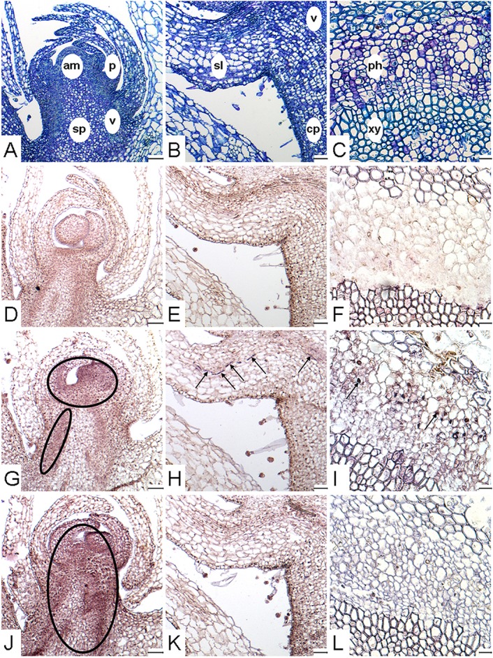
Figure 5.

Localization of PrSUT expression in P. ramosa. (A–L) Paraffin-embedded sections (10 μm thick) of parasitic tissues. (A,D,G,J) Longitudinal sections focused on the apical part of growing subterranean shoot (stage S.IV). (B,E,H,K) Longitudinal sections focused on a scale leaf in the basal part of subterranean shoots (stage S.IV). (C,F,I,L) Cross-sections focused on mature vascular tissues in the apical part of flowering shoots (stage FS.V). (A–C) Sections stained with TBO, showing the different tissues of shoot. (D–F) Section showing in situ hybridization signal obtained with PrSUT sense probes. The sense probe picture is representative for both probes. (G–I) Section showing in situ hybridization signal obtained with PrSUT1 antisense probe. (J–L) Section showing in situ hybridization signal obtained with PrSUT3 antisense probe. Open ovals and black arrows indicate positive hybridization signals. am, apical meristem; cp, cortical parenchyma; p, scale-leaf primordium; ph, phloem; sl, scale leave; sp, storage parenchyma; v, vasculature (immature vascular tissues); xy, xylem. Bars: (A,B,D,E,G,H,J,K): 100 μm; (C,F,I,L): 25 μm.
