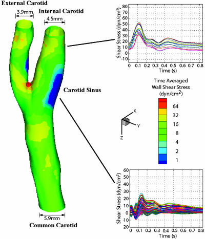
Fig. 1.
Hemodynamics in human carotid bifurcation manifests distinct site specificity. The left carotid anatomy of a normal human subject (27-year-old male) was reconstructed from noninvasive MRI measurements. The color-coded map shown in this carotid model displays the time-averaged shear stress magnitude at different points along the vascular wall. Regions in the distal internal carotid artery and carotid sinus were selected, and the time-dependent shear stress values along the mean flow direction from multiple individual points within each region were plotted for one cardiac cycle. For the carotid sinus, all of the finite element surface grid points within a 4-mm diameter circle in the sinus region (total 199 grid points) were chosen, and the time-dependent shear stresses along the mean flow direction from individual points were plotted (Lower Right). Each waveform depicted thus represents the time-dependent wall shear stress profile at a given point in each region of interest.

