Figure 1.
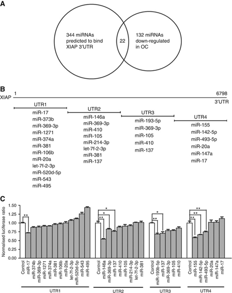
Screening for miRNAs targeting XIAP. (A) The Venn diagram shows the predicted XIAP 3′UTR binding miRNAs that were reported to be down-regulated in ovarian cancer. (B) Schematic diagram of the XIAP 3′UTR and miRNAs binding to the cognate fragments of XIAP 3′UTR. (C) Effects of 22 miRNAs on the activity of a luciferase reporter gene that is fused to four fragments of the XIAP 3′UTR in 293T cells using a dual-luciferase reporter assay. The luciferase activity of the transfected cells was measured 48 h post transfection. The results were presented as the relative luciferase activity and were normalised to the control, which was assigned a value of 1. The values represent the mean±s.d. from three independent transfection experiments. Significant differences from the control value are indicated by *P<0.05 and **P<0.01.

