
Figure 1.
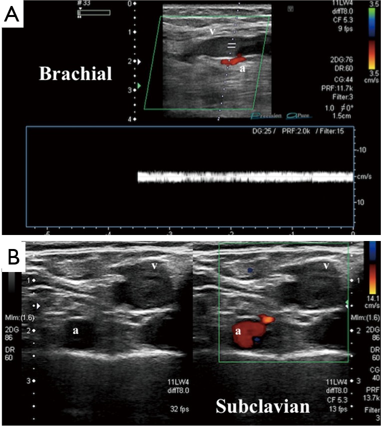
Compression ultrasonography showing an acute deep vein thrombosis (DVT) of the brachial and subclavian vein in cross-sectional and longitudinal views. (A) Longitudinal view of the brachial vein (v) showing hyperechogenicity and lack of color Doppler flow. Adjacent brachial artery (a) demonstrates antegrade flow; (B) cross-sectional view of the subclavian vein showing hyperechogenicity, lack of color Doppler flow, and lack of compression, consistent with acute DVT. Adjacent subclavian artery (a) demonstrates normal color Doppler flow.