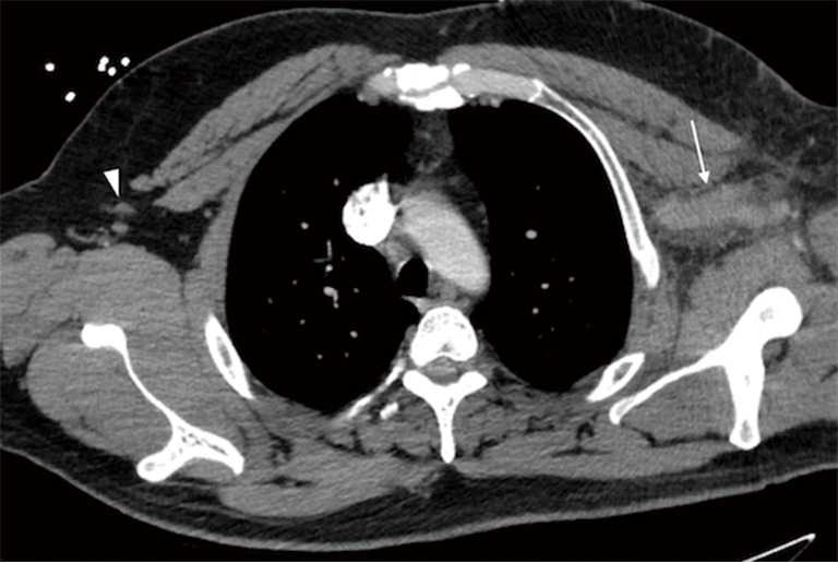
Figure 2.

Axial contrast-enhanced computed tomography (CT) of the chest showing an enlarged left axillary vein (arrow) with a large amount of surrounding fat stranding. The engorged vessel with surrounding inflammatory changes indicates an acute development of venous hypertension as a result of more central venous obstruction and thrombosis, in this case at the level of the thoracic inlet. The lack of collateralized vessels also suggests an acute presentation. The contralateral axillary vein (arrowhead) is shown for comparison.
