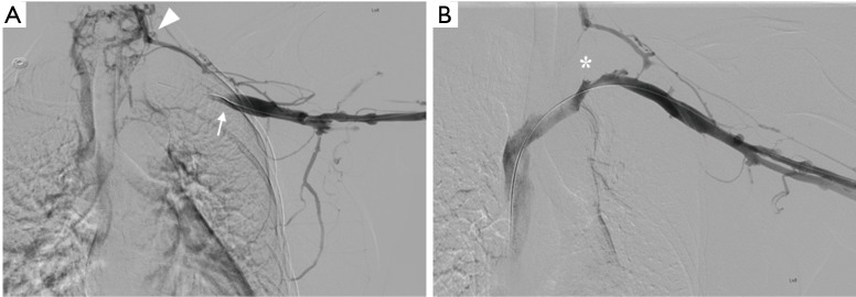
Figure 3.
Digital subtraction venography (DSV) of the left upper extremity and chest following local injection of iodinated contrast material through a sheath within the left brachial vein. (A) Prior to thrombolysis, there is abrupt termination of flow within the axillary vein at the level of the thoracic inlet (arrow), with redistribution of flow towards a multitude of collateral vessels within the neck (arrowhead); (B) following 24 hours of catheter-directed thrombolysis, there is restoration of flow within the subclavian and innominate veins extending to the superior vena cava. There is persistent stenosis (*) at the thoracic inlet, prompting referral to vascular surgery for decompression.

