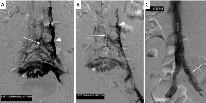
Figure 7.
Digital subtraction venography (DSV) of the left common iliac vein prior to and following stent placement for May-Thurner syndrome. (A,B) Transjugular venography prior to stent placement demonstrates abrupt termination of flow (arrow) within the left common iliac vein just prior to the inferior vena cava (IVC) confluence. A large ascending lumbar vein (arrowhead) is seen as well as deep pelvic varices (*). There is no flow within the IVC (location indicated by the descending transjugular venous catheter). Incidental note is made of a Greenfield IVC filter; (C) repeat venography following stent placement (arrow) shows restoration of patency to the left common iliac vein, with normal flow of contrast into the IVC and right common iliac vein.

