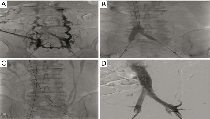
Figure 4.
Case #1 is a 58-year-old male with chronic thrombosis of the infrarenal IVC and bilateral common and external iliac veins. (A) Initial bilateral common iliac venogram demonstrates chronic occlusion of the bilateral common iliac veins and infrarenal IVC. Multiple bilateral venous collaterals are present; (B) fluoroscopic image of the pelvis demonstrates recanalization of the chronic occlusions of the bilateral iliac veins and infrarenal IVC. Angioplasty of the bilateral common iliac veins was performed; (C) fluoroscopic image of the abdomen demonstrates placement of Wallstents (Boston Scientific, Natick, MA, USA) within the IVC and bilateral common iliac veins. Incidentally noted is a duplicated left renal collecting system; (D) digital subtraction venography (DSV) demonstrates restored patency of the IVC and bilateral common iliac veins status post angioplasty and Wallstent placement.

