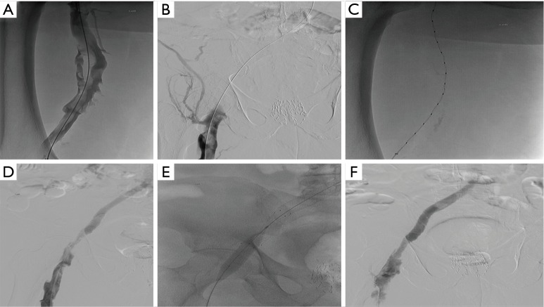
Figure 5.
Case #2 is a 73-year-old male with left lower extremity DVT and leg swelling for 5 weeks. (A) While in a prone position, a left femoral venogram demonstrates multiple filling defects consistent with thrombus; (B) digital subtraction venogram (DSV) of the left proximal femoral vein demonstrates multiple filling defects and complete occlusion of the left external and common iliac veins; (C) fluoroscopic image of an EKOS catheter (EKOS, a BTG International Group company, Bothell, WA, USA) placed into the left common femoral, left external iliac vein and left common iliac vein; (D) DSV of the left common femoral, left external iliac vein and left common iliac vein status post catheter directed thrombolysis via an EKOS catheter with improved venous blood return and decreased thrombus burden; (E,F) fluoroscopic and DSV images demonstrating angioplasty and placement of 14 cm × 6 cm Lifestent (Bard, Tempe, AZ, USA) in the left common iliac vein with markedly improved vascular patency of the left common femoral vein, left external iliac vein, and left common iliac vein. DVT, deep venous thrombosis.

