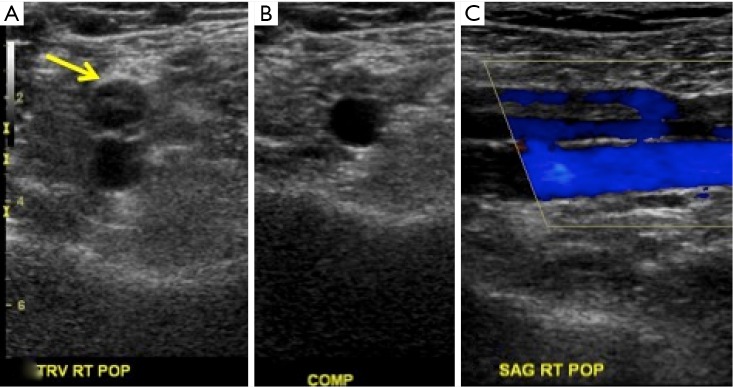
Figure 2.
Chronic recanalized deep vein thrombosis on ultrasound. Gray scale ultrasound examination of the right popliteal vein demonstrates echogenic venous wall and a compressible lumen with an eccentric linear area of echogenic material (arrow in A). Doppler flow is noted around this linear area of echogenic material.

