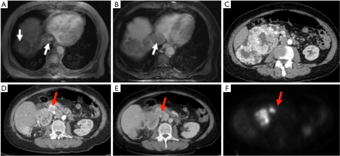
Figure 6.
Tumor thrombus. A patient with hepatocellular carcinoma shows a thrombus within IVC showing arterial enhancement (A) with washout on delayed phase (B) similar to the mass in the segment 7 of liver. Another patient with a large RCC arising from right kidney (C) shows a tumor thrombus in right renal vein (D,E; arrows) that shows FDG uptake on PET image (F).

