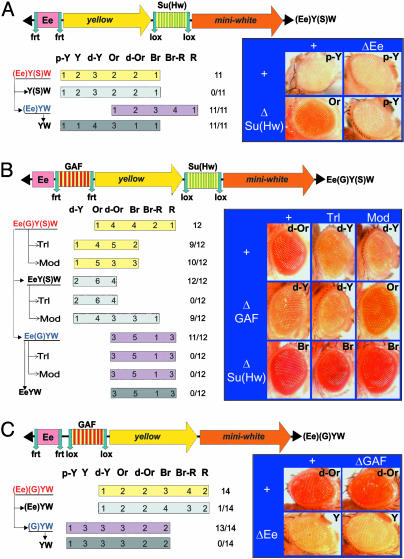
Fig. 3.
GAF-binding sites neutralize the enhancer-blocking activity of the Su(Hw) insulator. Transgenes are schematically represented above each panel. The transgenic lines are sorted according to their eye colors. Wild-type mini-white expression results in bright red eye color (R), whereas the absence of expression results in white eyes (W). Intermediate levels of pigmentation are defined by eye color ranging from pale yellow (p-Y) to yellow (Y), dark-yellow (d-Y), orange (Or), dark orange (d-Or), brown (Br), or brown-red (Br-R), reflecting increasing levels of miniwhite expression. The number of lines in each category is indicated. miniwhite expression levels were determined without excision as well as after excision of the functional elements flanked by loxP and frt sites. Analysis of miniwhite expression in a wild-type, TrlR85 heterozygous mutant and in a Mod(mdg4) homozygous mutant background (mod(mdg4)u1 and mod(mdg4)T6) is indicated (+, Trl, and Mod, respectively). The number of lines in which the eye pigmentation changes after excision of an element or in a mutant background, compared with the original lines, is indicated over the total number of lines. (A) Structure of the (Ee)Y(S)W transgene. Arrows indicate the excision of an element to produce the derivatives Y(S)W, (Ee)YW, and YW. Representative eye pictures are shown. (B) Structure of the Ee(G)Y(S)W transgene and eye colors of the Ee(G)Y(S)W lines and their derivatives. (C) Structure of the (Ee)(G)YW transgene and eye colors of the (Ee)(G)YW lines and their derivatives.

