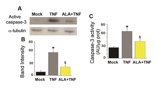
Fig. 1.

ALA reduces caspase-3 activity in C2C12 myoblasts. A) The representative Western bolt shows cleaved caspase-3 expression as assessed in C2C12 cells incubated for 48h in DM (differentiation medium) alone (Mock) or with TNF (tumor necrosis factor-α) or TNF and ALA (α-linolenic acid). α-tubulin was used as a loading control. B) Bands intensities of the western blot signals in (A). Values are expressed as arbitrary units. C) Bars are representative of caspase-3 activity measured by the fluorimetric assay. Fluorescence values, expressed as arbitrary units (AU), were normalized to protein content.
Data derived from three separate experiments are presented as the means±SD. * P<0.05 compared with Mock cells; § P<0.05 compared with the TNF group.
