
Fig 2.
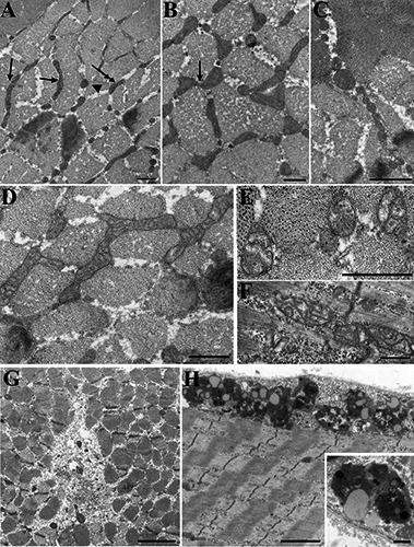
Shape and inner architecture of mitochondria are quite variable in MH+ fibers: evidence of some breakdown. A) Mitochondria in MH+ biopsies are mostly located in the I band as in MH-, but the elongated forms (arrows) are limited in length and in distribution. Short mitochondria segments (double arrow) and round profiles (arrowhead) are more frequent (Table 2). B) Shorter mitochondria are often aligned and sometimes apparently connected to each other (see also Fig. 2B). C) Very small round mitochondria forming a “chain” in the IM space are indicative of fragmentation. Such events are fairly frequent in MH+ fibres (Table 2). D-F) Mitochondrial internal architecture is altered in the majority of MH+ biopsies. Mild reduction of matrix density and cristae disarrangement (D) is detected in the majority of MH+ fibers. Highly disarranged cristae and almost empty matrix (E, F) are less frequent. G) Small unstructured regions containing some cellular debris, indicative of local degeneration are found in both MH- and MH+. The example is from an MH+ biopsy. H) Chain of lysosomes and lypofucsin granules at the periphery of the fibre and a detail in the inset. These represent residual bodies remaining after degradation of cytoplasmic components. The two examples are from MH- biopsies, but they are also frequent in MH+. Bars = A,B, D-F: 0.5 µm; C: 1 µm; G,H: 2 µm, insert 0.5 µm.