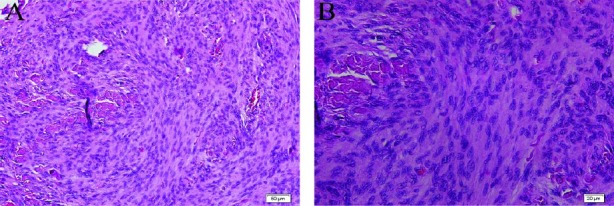
Figure 4.

Histopathology of specimen resected during surgery. H&E staining shows cells of tumor, composed of spindle cells arranging in whorl patterns (A). H&E staining shows ovaloid and spindle cells of tumor with plentiful cytoplasm and psammomatous calcification (B)
