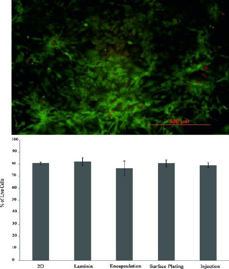
Figure 8.

Live/Dead fluorescence image of human meningioma stem-like cells injected in PM after 3 days of culture. Red fluorescent nuclei indicate dead cells and live cells exhibit green fluorescent. Quantification of the staining by manual counting of living and dead cells of 2D, laminin, and 3D cultures are shown. There was no difference among the groups in terms of cell viability after 3 days of culture
