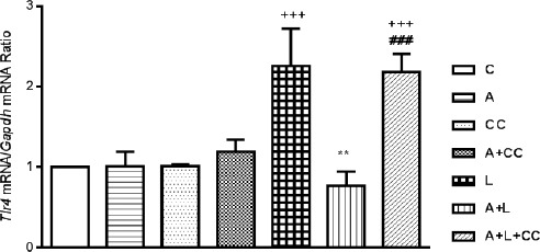
Figure 4.

Expression of Tlr-4 mRNA in mice myocardial tissues following 8 hr treatment, intraperitoneally, with A (A-769662 10 mg/kg), CC (compound-C 20 mg/kg), A+CC (A-769662 10 mg/kg+compound-C.20 mg/kg), L (LPS 2 mg/kg), A+L (A-769662 10 mg/kg+LPS 2 mg/kg) and A+L+CC (A-769662 10 mg/kg+2 mg/kg+compound-C 20 mg/kg) where C is control. mRNA was isolated and converted to cDNA. The expression of Tlr-4 mRNA was analyzed by real time PCR. Data are expressed as the mean±SEM of the four independent experiments. The significant of results was tested by a Pair Wise Fixed Reallocation Randomization Test using REST software. +++P<0.001 from respective control value; **P<0.01 as compared with the LPS group, ###P<0.001 as compared with the A+L group
