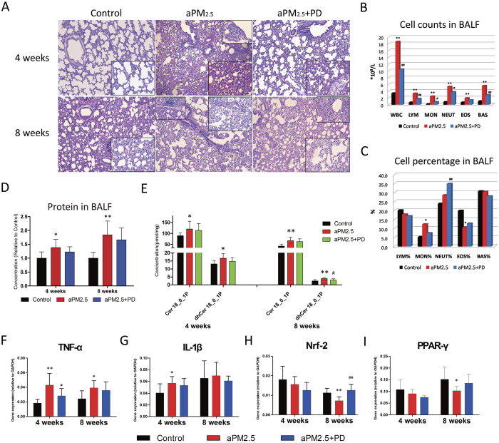
Figure 5. PD prevented aPM2.5-induced inflammation in the pulmonary interstitial space.
(A) A histological analysis of lung injury in rats. Representative low-power ( × 100) and high-power (inset, × 400) H&E-stained lung sections after exposure for 4 and 8 weeks. (B) Cell counts and (C) cell percentages in BALF after exposure for 8 weeks. The results are expressed as means (n = 8). (D) Protein level in BALF. The concentrations are shown as relative to the control. (E) The change in the level of ceramides in the rat lung tissues. The results are expressed as means ± SD (n = 8). Relative gene expression of (F) tumor necrosis factor (TNF)-α, (G) interleukin (IL)1-β, (H) nuclear factor NF-E2-related factor 2 (Nrf2) and (I) peroxisome proliferator–activated receptor (PPAR-γ). Values are reported using the 2−ΔCt method. Samples were normalized to GAPDH gene expression. The results are expressed as means ± SD (n = 8). *P < 0.05 and **P < 0.01, versus the control group; #P < 0.05 and ##P < 0.01, versus the aPM2.5 group.

