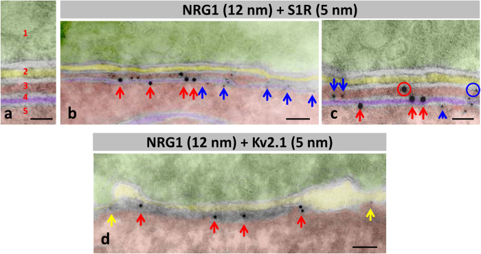
Figure 3. Immunolabelling using nanogold particles of ultrathin cryosection for the ultrastructural localisation of NRG1, S1R and Kv2.1 channels at C-boutons.
To facilitate the localisation of C-boutons, the analysis was performed in hypoglossal MNs. A detail of the organization of compartments at C-bouton synapses in negatively stained cryosections is depicted in (a) 1 = presynaptic (green), 2 = intersynaptic extracellular space (yellow), 3 = postsynaptic cytoplasmic compartment lodged between postsynaptic membrane and subsynaptic cistern (SSC, red), 4 = SSC (violet), and 5 = MN cytoplasm (red). The same colour code is used in (b–d). (b,c) Double immunolabelling for NRG1 and SR1; NRG1 (red arrows) is mainly associated with SSC forming a cluster segregated from SR1 (blue arrows). (c) A minor proportion of gold particles (encircled) are located at the postsynaptic membrane. (d) nanogold particles labelling Kv2.1 are located on the periphery (yellow arrows) of C-bouton enriched with a NRG1 cluster (red arrows). Scale bar: in (a,c) = 25 nm; in (b,d) = 50 nm.

