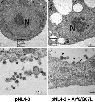
Fig. 5.
HIV-1 particles assemble in Arf6/Q67L-induced vesicles. HeLa cells transfected with pNL4-3 alone (A and C) or cotransfected with pNL4-3 and the HA-tagged Arf6/Q67L expression plasmid (B and D) were observed by transmission electron microscopy (EM). Higher magnification of boxed areas in A and B is shown in C and D, respectively. Note that virus particles as well as budding structures are detected on the membrane of Arf6/Q67L-induced vesicles (arrows in D). In the cultures cotransfected with pNL4-3 and the Arf6/Q67L expression plasmid, 42% of the virus particles were detected in association with these vesicles, whereas only 6% of virions in cultures transfected with pNL4-3 alone was associated with intracellular vesicles. Scale bars are shown in each panel. Nuclei are indicated (N).

