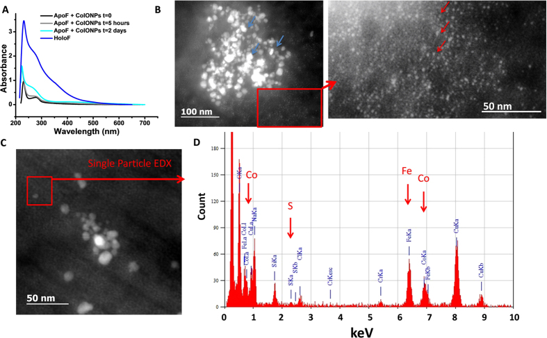
Figure 5. Metal transfer from NPs to apoferritin proteins.
(A) UV-vis spectra of ApoF incubated with CoIONPs in acidic medium for 5 hours and 2 days, in comparison to ApoF and HoloF. The growth of absorbance shoulder at 280 nm indicates metal filling of the protein. (B) STEM-HAADF images of CoIONPs (blue arrows) incubated with ApoF for 2 months. Metal-filled proteins appear as small particles (red arrows). (C,D) Single particle EDX analysis of the red-contoured area in C confirms occupancy of iron, cobalt and sulfur in single ApoF with relative atomic percentages of 64.7%, 28.2% and 7.13% respectively.

