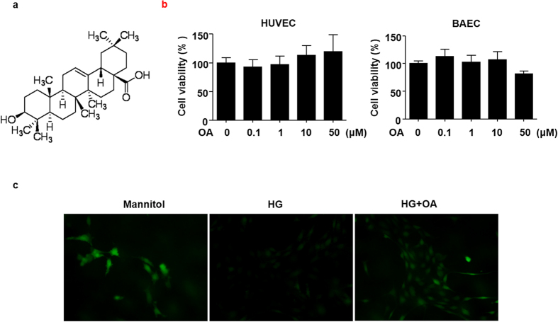
Figure 1. OA improved high glucose-induced NO reduction in BAECs.
(a) The chemical structure of OA. (b) HUVECs and BAECs were treated with indicated concentrations of OA for 24 h, and cell viability was measured by MTT. All data were expressed as mean ± SEM of triplicate experiments. (c) BAECs were preincubated with or without OA (10 μM) for 12 h, then, treated with high glucose (HG, 30 mM, 12 h), mannitol served as vehicle control to HG. NO was detected by using DAF-FM diacetate (40 × objective).

