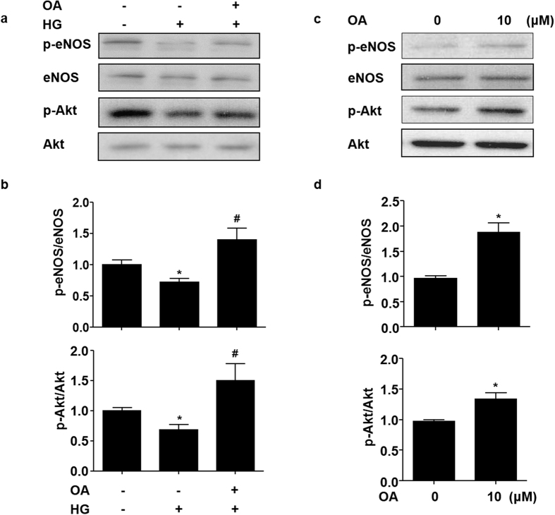
Figure 2. OA attenuated the high glucose-induced impairment of Akt-Ser473 and eNOS-Ser1177 phosphorylation.
(a) HUVECs were pretreated with or without OA (10 μM) for 12 h, and then exposed to normal (5 mM) or high (30 mM) glucose for 12 h. Protein levels of p-eNOS, eNOS, p-Akt and Akt were detected by using western blotting. (b) Quantification of p-eNOS/eNOS and p-Akt/Akt. (c) HUVECs were stimulated with OA (10 μM) for 24 h, cell lysates were analyzed to determine p-eNOS, eNOS, p-Akt and Akt protein levels by using western blot. (d) Quantification of p-eNOS/eNOS and p-Akt/Akt levels in HUVECs. Data were shown as mean ± SEM of independent experiments. *P < 0.05 vs. vehicle control. #P < 0.05 vs. HG.

