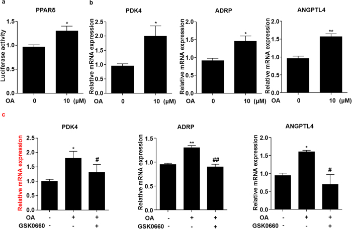
Figure 3. OA activated PPARδ in ECs.
(a) BAECs were transfected with PPRE-luc and PPARδ plasmids and then treated with OA (10 μM) for 24 h. The luciferase activities were shown as fold changes in relation to the control. (b) HUVECs were stimulated with OA (10 μM) for 24 h, PDK4, ADRP and ANGPTL4 mRNA levels were assessed by using qRT-PCR. (c) HUVECs were pretreated with GSK0660 (1 μM) for 1 h, then exposed to OA (10 μM) for 24 h, mRNA levels of PDK4, ADRP and ANGPTL4 were assessed. *P < 0.05, **P < 0.01 vs. vehicle control. #P < 0.05, ##P < 0.01 vs. OA.

