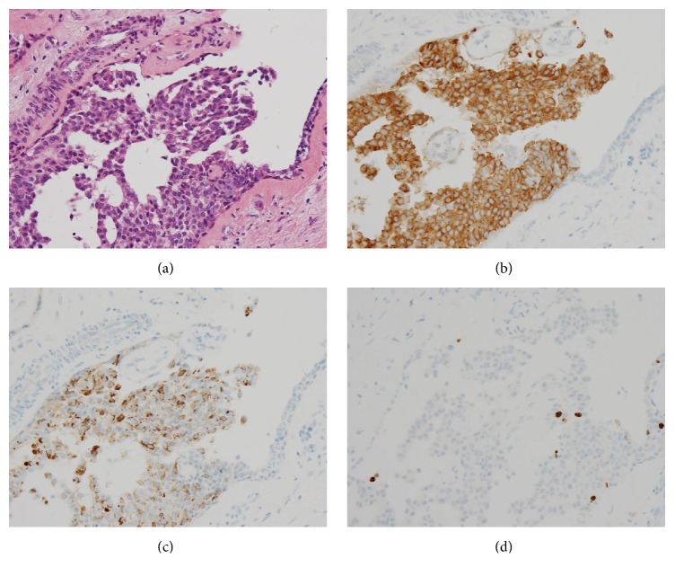
Figure 5.
(a) The tumor cells show a solid papillary growth pattern (HE stain, original magnification ×400). (b) The tumor cells show positive immunoreactivity for synaptophysin (Immunohistochemistry, original magnification ×400). (c) The tumor cells show positive immunoreactivity for chromogranin A (Immunohistochemistry, original magnification ×400). (d) A few tumor cells show positive immunoreactivity for Ki-67 (Immunohistochemistry, original magnification ×400).

