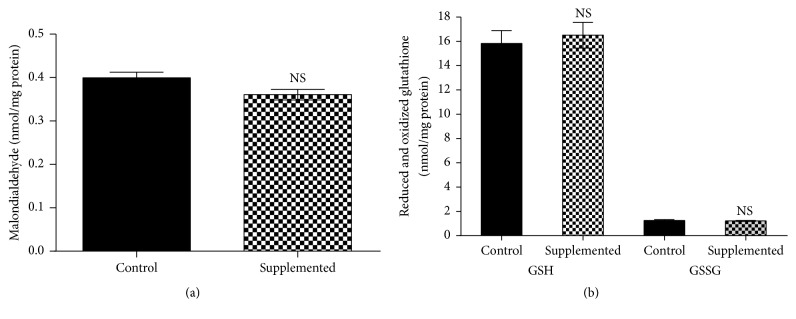
Figure 1.
Effect of biotin supplementation on liver malondialdehyde and reduced and oxidized glutathione in mice fed a biotin-supplemented diet for 8 weeks. (a) Malondialdehyde concentrations. Values represent the mean ± SEM of 5 mice from each group. (b) Reduced and oxidized glutathione. Black bars: control group; patterned bars: biotin-supplemented group. Values represent the mean ± SEM of 8 mice from each group. All measurements were performed in triplicate. NS p > 0.050.

