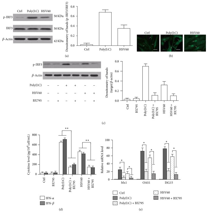
Figure 6.
Poly(I:C) and HSV60-induced immune responses through IRF3 activation. (a) Phosphorylation of IRF3 in hAD-MSCs. hAD-MSCs were stimulated with poly(I:C) or HSV60 for 2 h. Cell lysates were analyzed by Western blot to probe phospho-IRF3 (p-IRF3) and total IRF3. β-Actin was used as loading control. (b) Nuclear translocation of IRF3. hAD-MSCs were stimulated with poly(I:C) (middle panel) or HSV60 for 4 h. Immunofluorescence staining was performed by IRF3 antibody. Arrows show the representatives of nucleus in hAD-MSCs. (c) Inhibition of IRF3 activation. hAD-MSCs were stimulated with poly(I:C) or HSV60 alone or with poly(I:C) or HSV60 after 2 h before incubation with 1 μg/mL BX795 (an inhibitor of IRF3 activation). At 2 h after poly(I:C) or HSV60 stimulation, p-IRF3 and total IRF3 level were determined by Western blot. (d) IFN-α and IFN-β secretion. The cells were treated as (c). At 24 h after poly(I:C) or HSV60 stimulation, the cytokine levels in media were measured using ELISA. (e) Expression of antivirus proteins. The cells were treated as (c). At 6 h after poly(I:C) or HSV60 stimulation, the mRNA levels of antiviral proteins, ISG15, OAS1, and Mx1, were determined using real-time PCR. The cells that were treated with LyoVec alone were served as Ctrl. Images represent at least three experiments. Scale bar = 20 μm. Data are the means ± SEM of three experiments. ∗ P < 0.05; ∗∗ P < 0.01.

