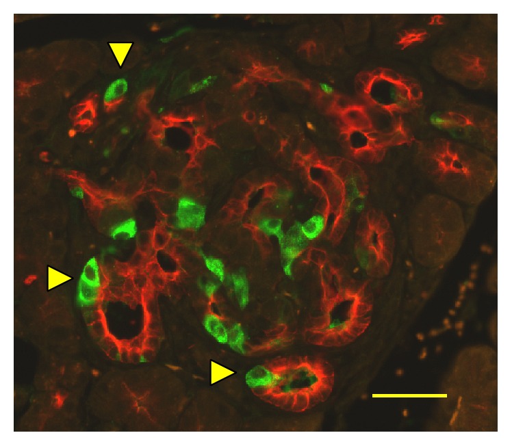
Figure 5.

Insulin-expressing cells in the Tg mice ductal structures. Pancreas sections from Tg mice at 40 weeks of age costained with antibodies to insulin (green) and cytokeratin 17/19 (red). Yellow triangles denote insulin-positive cells in the structure. Bar, 40 μm.
