Figure 4.
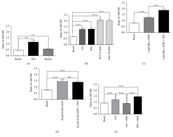
Effects of SPA on cytosolic calcium concentrations. Cytosolic calcium variations were measured using the Fura-2AM in the mouse MIN6-B1 β-cell line. Fura2-AM absorbance ratio (340/380) was given for the time point with the maximal signal. (a) At low glucose concentration (5 mM), SPA (10−7 M) induced a significant cytosolic calcium rise (n = 18, p < 0.01) that did not return to basal level after washing out (p < 0.01). (b) Comparing SPA and ex4 effects, controls were performed using either KCl (25 mM) or glucose (20 mM) (n = 17). (c) Preincubation of MIN6-B1 cells with 5 mM 8Br-cAMP did not prevent the stimulating effect of SPA on calcium rise (n = 48). (d) When MIN6-B1 cells were preincubated with 50 mM 8Br-cAMP, SPA was not able to increase intracellular calcium (n = 12). (e) Preincubation of MIN6-B1 cells in the presence of 1 μM H89 significantly inhibited the ex4 effect but not that of SPA (n = 38). n indicates the number of responding cells in each of three experiments. Results are expressed as mean ± SEM; ∗∗p < 0.01, ∗∗∗p < 0.001, and ns: nonsignificant.

