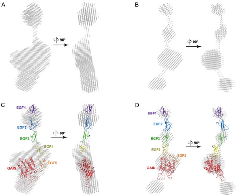
Figure 4.
PNGase F deglycosylation of CD97ECD caused shape change in SAXS measurement. (A) SAXS envelope of His-CD97ECD WT protein shape. (B) SAXS envelope of PNGase F digested His-CD97ECD WT protein shape. (C) Docking results of CD97ECD homology models to SAXS envelope of His-CD97ECD WT. (D) Docking results of CD97ECD homology models to SAXS protein shape of PNGase F digested His-CD97ECD WT.

