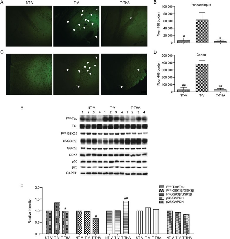
Figure 10.
THA reduced Tau phosphorylation by inhibiting GSK3β activity in the APP/PS1 transgenic mice. (A–D) Immunohistochemical assay was performed to detect the Tau phosphorylation burden in the hippocampus (A) and cortex (C); white arrows indicate Tau phosphorylation. (B) Statistical analysis of (A). (D) Statistical analysis of (C). Scale bar, 100 μm. (E) The levels of Tau at Serine396, GSK-3β (phosphorylated at Ser9 or Tyr216), GSK-3β, CDK5 and p35/p25 were tested by Western blot. (F) Densitometry analysis of (E). GAPDH was used as a loading control in the Western blot assays. NT-V, non-transgenic vehicle group; T-V, transgenic vehicle group; T-THA, transgenic mice administration with 300 mg THA/kg per day group. Values are the mean±SEM. t test. #P<0.05, ##P<0.01 compared with T-V.

