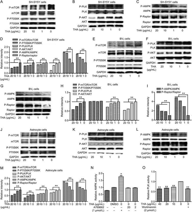
Figure 5.
Inhibition of PI3K/AKT and activation of the AMPK/Raptor pathway were involved in promotion of THA-mediated Aβ clearance. (A–M) Cells were cultured with different concentrations of THA (20, 10, 1, or 0 μg/mL) for 24 h, and the levels of P-mTOR, mTOR, P-P70S6K, P70S6K, P-PI3K, PI3K, P-AKT, AKT, P-AMPK, AMPK, P-Raptor, and Raptor were detected by Western blot in SH-SY5Y cells (A–C), BV2 cells (E–G) and astrocytes (J–L). (D) Densitometry analysis of Figures (A–C). (H) Densitometry analysis of Figure (E and F). (I) Densitometry analysis of Figure (G). (M) Densitometry analysis of Figures (J–L). (N) An AMPK enzyme reaction system was directly incubated with different concentrations of THA (20 or 2 μg/mL), and the in vitro AMPK activity was then evaluated. A769662: AMPK activator56. (O) A PI3K enzyme reaction system was directly incubated with different concentrations of THA (40, 20, 10, or 0 μg/mL), and the in vitro PI3K activity was then evaluated. Wortmannin: PI3K inhibitor55; GAPDH was used as a loading control in the Western blot assays. The results were obtained from three independent experiments. Values are the mean±SEM, one-way ANOVA, Bonferroni's multiple comparison test. n=3. *P<0.05, **P<0.01 compared with the control group, control group: 0 μg/mL THA treatment group.

