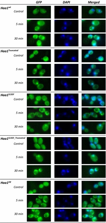
Fig. 4.

Subcellular localization of Haa1 before and after exposure of the cells to acetic acid. Results are shown for the wild-type CEN.PK113-7D strain (Haa1WT) and the Haa1S135F_Truncated, Haa1Truncated, Haa1S135F and Haa1OE CEN.PK113-7D strains. Haa1 localization was determined in cells immediately before and 5 and 30 min after the transfer to medium containing 50 mM acetic acid (pH 4.5). Cells were stained with 4′,6-diamidino-2-phenylindole (DAPI) to label nuclear DNA. Visualization was performed using fluorescence microscopy to detect DAPI and GFP fluorescence signals. ImageJ software was used to overlay the images
