
Fig. 2.
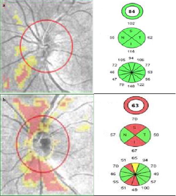
Retinal Nerve Fiber Layer (RNFL) thickness analysis using optic disc cube 200 × 200 feature depicting on RNFL thickness deviation map a left eye of patient with non-proliferative diabetic retinopathy showing RNFL thinning, b left eye of patient with proliferative diabetic retinopathy showing thinning of RNFL