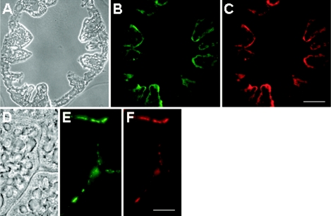
FIG. 4.
Thin sections of the midgut glands of P. scaber, hybridized with fluorescently labeled oligonucleotide probes. The photomicrographs show the same microscopic fields of a transverse section (A to C) and a tangential section (D to F), viewed with phase-contrast microscopy (A and D) and with epifluorescence microscopy by using filter sets for probe EUB338 (B and E), which is specific for Bacteria, and for probe PsSym352 (C and F), a group-specific probe. (C) Scale bar = 100 μm. (F) Scale bar = 10 μm.

