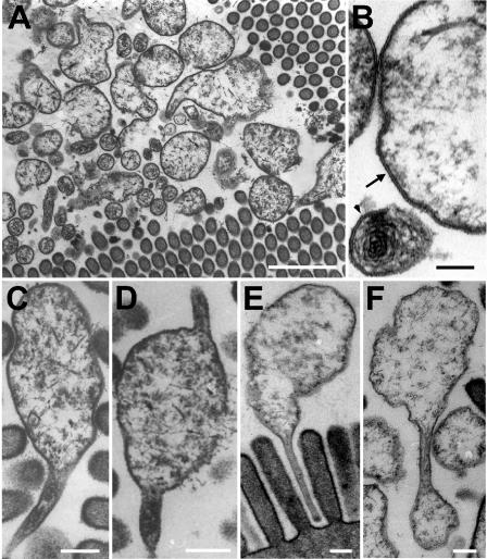
FIG. 5.
Transmission electron micrographs of bacterial symbionts in the midgut glands of P. scaber, showing large and small spherical cells in close association with the microvilli (A); both large and small cells lacking a cell wall and bound by a single unit membrane, indicated by an arrow (large cell) and an arrowhead (small cell) (B); the frequently observed stalk on one cell pole (C), on both cell poles (D), and inserted into the gaps between the microvilli of the brush border (E); and cell division by budding from the stalks (F). (A) Scale bar = 1 μm; (B) Scale bar = 0.1 μm; (C to F) Scale bars = 0.2 μm.

