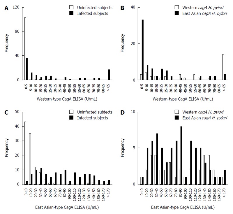
Figure 3.

The distribution of CagA antibody ELISA titers is dependent on Helicobacter pylori infection status and Helicobacter pylori CagA-type. A: Histogram of Western-type CagA ELISA titers of Helicobacter pylori (H. pylori)-infected (gray bars) and uninfected subjects (white bars); B: Histogram of Western-type CagA ELISA titers of H. pylori infected subjects is dependent on cagA genotype; Western-type cagA H. pylori (white bars), East Asian-type cagA H. pylori (gray bars); C: Histogram of East Asian-type CagA ELISA titers of H. pylori infection and uninfected subjects. Bar coloring is the same as in (A); D: Histogram of East Asian-type CagA ELISA titers of H. pylori infected subjects is dependent on cagA genotype. Bar coloring is the same as in (B).
