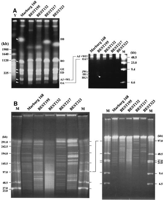
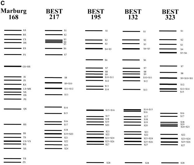
FIG. 3.
Identification of I-CeuI and SfiI fragments from B. subtilis (natto) strains. (A) I-CeuI digests of genomic DNA of four B. subtilis (natto) strains. I-CeuI digests of the strains indicated were electrophoresed. Lanes Y and M contained yeast genome DNA and lambda phage DNA, respectively, as markers; sizes are indicated on the left and the right. I-CeuI fragments of the Marburg 168 genomic DNA designated in reference 44 are indicated by arrowheads. The fragment-like band in BEST195 indicated by an arrowhead was an artifact because it was not reproduced in separate running conditions. The running conditions for the gel on the left were 3 V/cm, a pulse time of 8 min, and a running time of 44 h. Resolution of the I-CeuI fragments smaller than 150 kb is shown in the gel on the right, for which the running conditions were as follows: 3 V/cm, a pulse time of 3 min, and a running time of 32 h. (B) SfiI digests of four B. subtilis (natto) strains. SfiI fragments of different strains were resolved by pulsed-field gel electrophoresis with the following running conditions: 3 V/cm, a pulse time of 24 s, and a running time of 56 h (left panel) and 3 V/cm, a pulse time of 6 s, and a running time of 24 h (right panel). Resolution of the SfiI restriction fragments smaller than 100 kb is shown in the right panel. Lanes M contained lambda phage DNA as a size marker. (C) Schematic diagram of the SfiI fragments of the four B. subtilis (natto) strains. All the SfiI fragments determined experimentally are included. Sizes are shown in Table 2.


