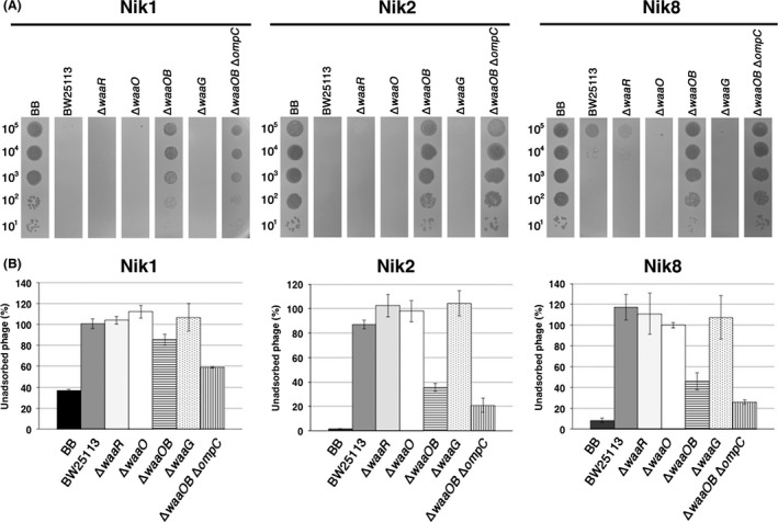
Figure 6.

Characterization of T4 Nik mutants. (A) The solution containing the number of phage particles (Nik1, left; Nik2, middle; Nik8, right panel) indicated on the left was spotted on a lawn of the E. coli strain indicated at the top and the plates were incubated at 37°C overnight. (B) Adsorption analyses of Nik1 (left), Nik2 (middle), or Nik8 (right) phages with the E. coli strain indicated at the bottom were performed. The relative numbers of unadsorbed phage particles at 10 min after phage addition were calculated with the numbers of input phage particles set to 100%.
