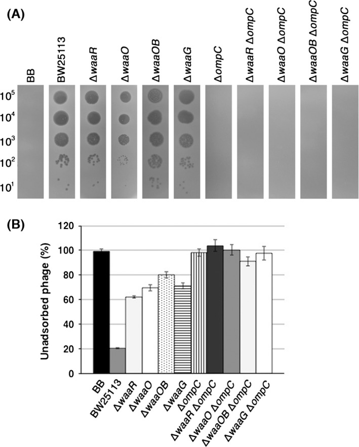
Figure 7.

Characterization of the T4 Nib mutant. (A) A solution containing the number of Nib phage particles indicated on the left was spotted on a lawn of the E. coli strain indicated at the top and the plates were incubated at 37°C overnight. (B) Adsorption analyses with the E. coli strains indicated at the bottom were performed as described in Experimental Procedures.
