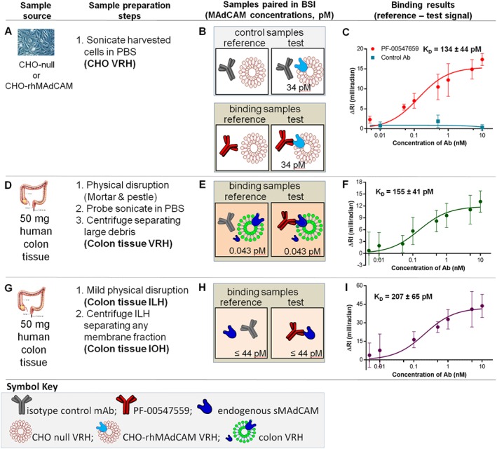
Figure 4.

mMAdCAM‐binding assays – thermodynamic KD in CHO cell VRH, human colon VRH and tissue IOH. (A) CHO‐WT VRH and CHO‐rhMAdCAM VRH are prepared by sonication in PBS, (B) binding interaction samples are paired and (C) saturation binding isotherm used to quantify the thermodynamic KD{CHO,mMAdCAM} of PF‐00547659 to mMAdCAM expressed in CHO cells. (D) Human (UC) colon tissue physically disrupted, sonicated and centrifuged to produce tissue VRH sample, (E) binding interaction samples are paired and (F) saturation binding isotherm used to quantify the thermodynamic KD{issue,mMAdCAM} of PF‐00547659 to tissue mMAdCAM in tissue. (G) Human (UC) colon tissue mildly physically disrupted and centrifuged to produce tissue IOH sample, (H) binding interaction samples are paired with (I) saturation binding isotherm used to quantify the thermodynamic KD{tissue,sMAdCAM} of PF‐00547659 to sMAdCAM in tissue. Error bars for all plots represent the standard deviations of replicate trials over five consecutive experiments performed on different days (n = 4–5).
