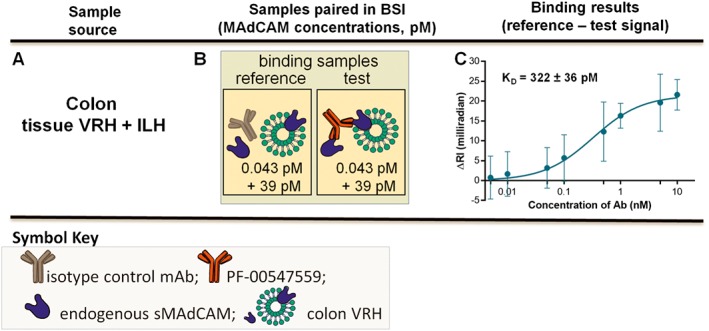
Figure 5.

Binding assays combining VRH and ILH – KD,app‐int in tissue VRH and ILH. (A) Tissue VRH is mixed with tissue ILH, (B) binding interaction samples are paired and (C) saturation binding isotherm used to quantify the KD,app‐int{issueVRH + 87.5%ILH} of PF‐00547659 to tissue mMAdCAM and tissue sMAdCAM. Error bars for all plots represent the standard deviations of replicate trials over five consecutive experiments performed on different days (n = 5).
