
Figure 1.
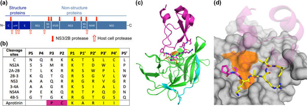
Design of aprotinin constructs mimicking dengue protease substrates. (a) Dengue virus polyprotein cleavage sites. (b) Polyprotein cleavage site sequences of DENV3 protease. (c) Aprotinin–DENV3 protease complex structure (3u1j). NS3 protease domain is in green, NS2B cyan, and aprotinin purple.10 (d) The binding loop of aprotinin is displayed as sticks and the residues screened with corresponding P1–P4′ substrate sequences are colored yellow.