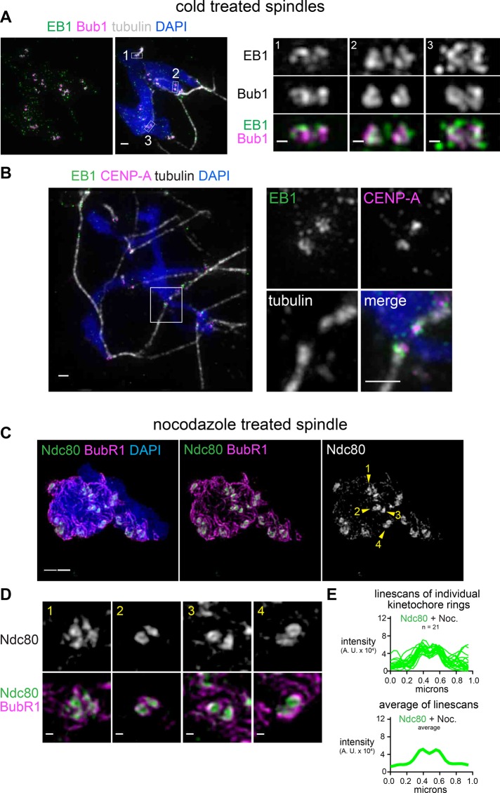
FIGURE 3:
Kinetochore rings may engage a hollow bundle of microtubules but do not depend on microtubules. (A, B) Spindles assembled in the presence of Alexa 488–labeled EB1 and incubated on ice for 2 min (scale bar, 1 μm). Pairs of sister kinetochores are shown at higher magnification (scale bars, 0.2 μm), with locations indicated by white boxes. (C, D) Mitotic chromosomes assembled in extract treated with 33 mM nocodazole to depolymerize all microtubules (scale bar, 1 μm). Pairs of sister kinetochores are shown at higher magnification (scale bars, 0.2 μm), with locations indicated by arrowheads. All images are maximum-intensity projections of SIM data. (E) Lines scans of ring-shaped Ndc80 signals in nocodazole-treated extract. Top, overlays; bottom, average intensity.

