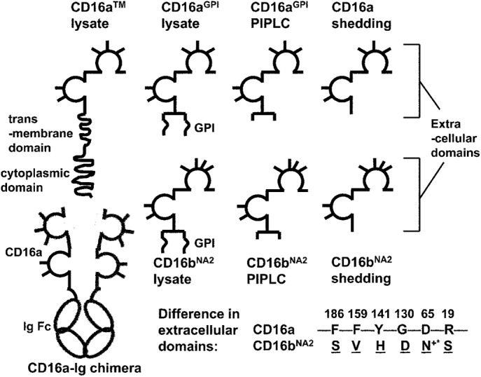
FIGURE 1:

Schematics of solubilized CD16 isoforms with different anchor structures and soluble CD16a–Ig chimera. The extracellular domains of CD16 are depicted as two Ig-like globules, with the N-glycosylation sites shown as sticks. An additional glycan is part of the GPI anchor, which acts as a linker between the C-terminus of the peptide and the phosphatidylinositol group. The amino acids in extracellular domains that differ between CD16a and CD16bNA2 are listed. +*The gained glycosylation site in CD16bNA2 due to the change D65 → N65.
