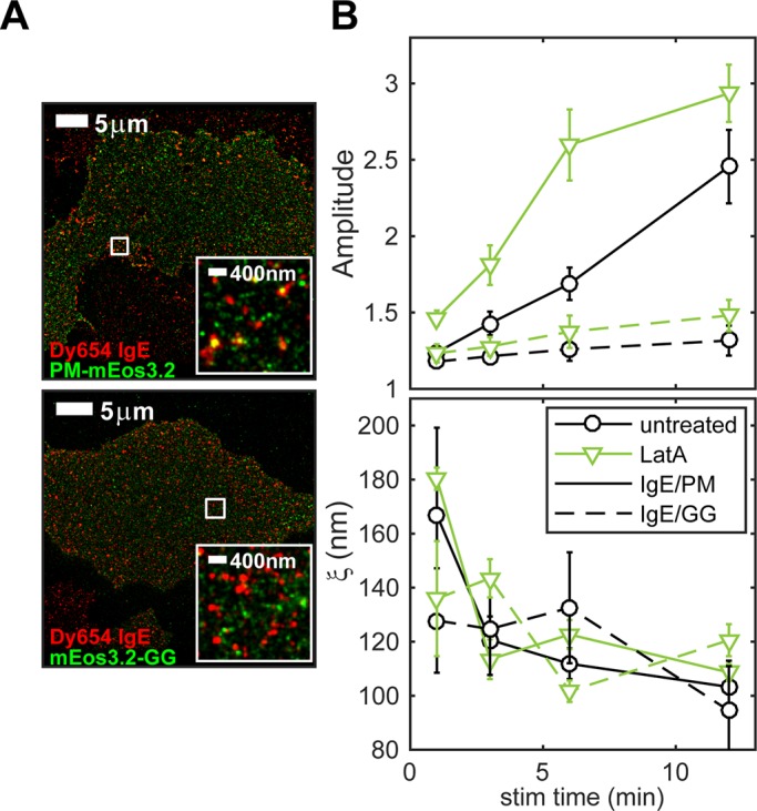
FIGURE 5:

Lipid anchorage of mEos3.2 constructs determines antigen-induced recruitment to IgE/FcεRI and the effects of cytoskeletal perturbation. (A) Representative two-color images of cells expressing PM-mEos3.2 (top) or mEos3.2-GG (bottom) sensitized with Dy654 IgE and stimulated for 6 min. (B) Average PM/IgE-FcεRI and GG/IgE-FcεRI cross-correlation fit parameters A and ξ as a function of stimulation time in RBL-2H3 cells. Cells were stimulated for 1, 3, 6, or 12 min with and without treatment with 1 μM latrunculin. Fit parameters are averaged over multiple cells for each time point (seven cells per time point for cells expressing both constructs and for both treatment conditions). Error bars denote SEM.
