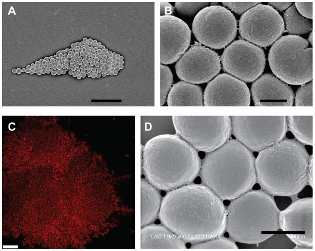
Fig. 2.
Images of S. aureus clumps. Washed cells of USA300 strain LAC were incubated with 18.5 μg/ml human fibrinogen and imaged using scanning electron microscopy (A, B, D) or confocal laser scanning microscopy (C). In panel C the cells were expressing DsRed from a plasmid. Scale bars represent 5 μm (A), 0.5 μm (B), 10 μM (C), and 0.5 μm (D).

