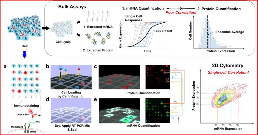
Figure 1.
Schematic diagrams of simultaneous quantification of mRNA and protein for single-cell level analysis. Conventional analytical methods for individual mRNA and protein quantification rely on bulk assays, with which either population-based analysis or correlation between mRNA and protein cannot be studied at the single-cell level. Our strategy for single-cell-level analysis and correlation studies utilizes a microwell device for single-cell loading and analysis. Individual cells stained with immunofluorescence antibody for the target protein were loaded into single wells on the microwell device, followed by imaging of the protein fluorescence intensities from the immunostained cells. The cells on the device were then directly lysed, and their target transcripts were amplified and tagged with a fluorescent probe via RT-PCR. Each window of the microwell device was imaged, and mRNA fluorescence intensities were extracted. The extracted fluorescence signals from protein and mRNA were analyzed and displayed for correlation using MATLAB software.

