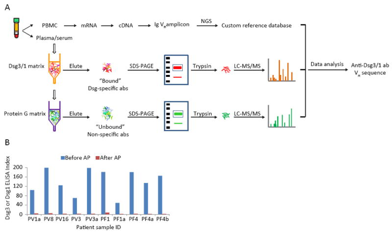
Figure 1. Proteomic platform to identify circulating pemphigus anti-Dsg abs.

(A) IgG heavy chains from Dsg-binding abs and from abs which do not bind to Dsg are analyzed by LC-MS/MS. Resultant spectra are searched against a custom database of all VH sequences from the same patient to identify ab peptides. Informative peptides match H-CDR3 AA sequences that define clonotypes of abs. (B) ELISA assays of PV or PF sera before and after affinity chromatography show that anti-Dsg ab is depleted by AP. PBMC, peripheral blood mononuclear cells. AP, affinity purification.
