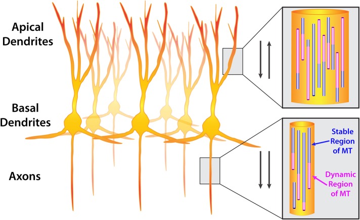
FIGURE 1:
Orientation of microtubules in CNS neurons. A group of neurons (cortical or hippocampal) showing apical dendrites, basal dendrites, and axons. Insets show microtubule orientation in dendrites and axons. Microtubules are composed of stable (purple) and dynamic (pink) regions. Dynamic regions undergo polymerization and depolymerization, termed dynamic instability. Arrows indicate that microtubules are oriented antiparallel in dendrites (plus and minus ends distal) and parallel in axons (plus ends distal).

